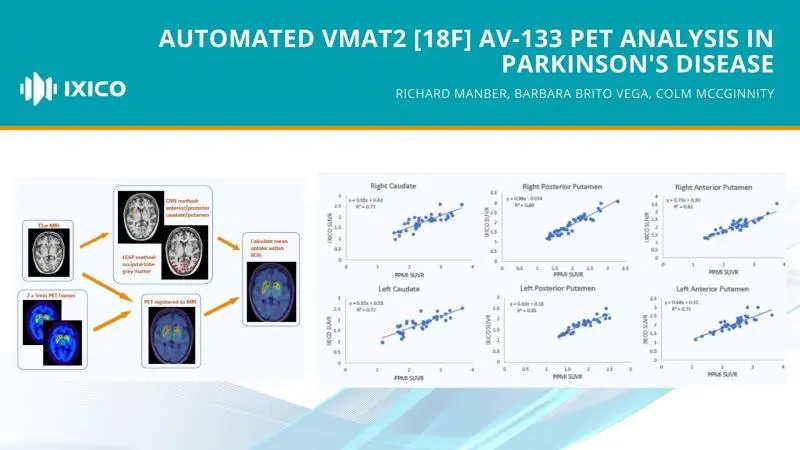
We discuss our latest work to evaluate a fully automated image analysis pipeline to process vesicular monoamine transporters type 2 (VMAT2) [18F]AV-133 tracer positron emission tomography (PET) images, by comparison with a methodology requiring manual intervention.
Our methods examined 41 PET scans from 21 idiopathic Parkinson's disease patients, alongside associated T1-weighted MRI images from the PPMI database. The PET images were registered to the MRI scans, and six brain regions were segmented using multi-atlas and convolutional neural network techniques. The Standardized Uptake Value Ratio (SUVR) was computed using the occipital grey matter as reference and then compared to PPMI's published results, assessing the correlation between the two datasets
Our fully automated pipeline to quantify VMAT2 [18F]AV-133 PET images produced comparable results to the PPMI pipeline requiring manual intervention, despite the differences in the definition of reference region (inclusion of white matter versus grey-matter only) and target regions (spherical VOI versus whole region ROI).
The proposed pipeline is objective and reproducible, therefore allowing for application in PD studies or other studies using nigrostriatal pathway integrity as a biomarker.