IXICO research presented at ESMRMB 2026 compares two MRI approaches for visualising the substantia nigra, a brain region strongly implicated in Parkinson's disease.
Download the poster
The study compared two neuromelanin-sensitive MRI acqusition techniques, MT-GRE and TSE, in healthy volunteers and introduced a robust, segmentation-free framework to quantitatively measure substantia nigra contrast. Both techniques successfully visualised the region, but MT-GRE provided consistently higher contrast between the substantia nigra and surrounding tissue than TSE.
The findings support the optimisation and more reproducible assessment of neuromelanin-MRI for future Parkinson's disease research and clinical trials. Further studies will evaluate the method's ability to differentiate patient groups and sensitively track disease-related changes over time.
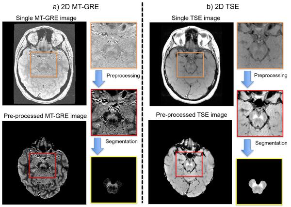
Representative MT-GRE (left) and TSE (right) images. Input panels show one of five scans; processed panels combine all five. Bottom panels are LEAP-derived midbrain-masked images, not SN segmentations.