Resources
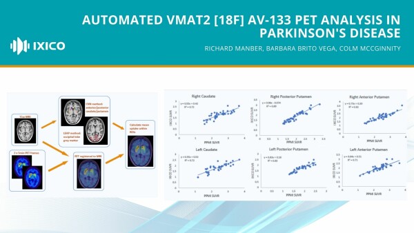
Automated VMAT2 [18F] AV-133 PET analysis in Parkinson's disease
We discuss our latest work to evaluate a fully automated image analysis pipeline to process vesicular monoamine transporters type 2 (VMAT2) [18F]AV-133 tracer positron emission tomography (PET) images, by comparison with a methodology requiring manual intervention.

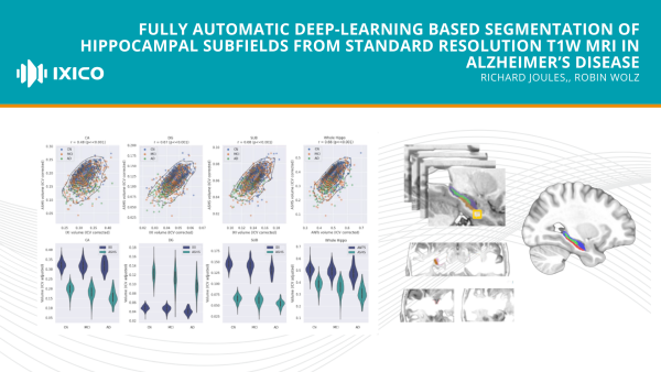
Fully automatic deep-learning based segmentation of hippocampal subfields from standard resolution T1W MRI in Alzheimer’s disease
Here we train an AI method to segment the hippocampus into subfields (CA1-3 CA4+DG, and Subiculum) from standard resolution T1W MRI alone to assess utility as biomarkers compared to whole hippocampal volume, in the absence of a high-resolution T2 MRI.

Huntington’s Disease Image Harmonization Consortium secures new member and funding to complete large-scale analysis of brain changes using IXIQ.Ai
New York, NY / London, UK, July, 12th 2023; The Huntington’s Disease Imaging Harmonization (HD-IH) Consortium, founded last year to conduct an unprecedented harmonization analysis of more than 6,000 participant-visit magnetic resonance images (MRIs) acquired from over 2,000 research participants, has now completed the initial phase and secured the necessary funding commitment to complete the…
MR Imaging in Ataxias Consensus Recommendations by the Ataxia Global Initiative Working Group on MRI Biomarkers
As members of the Ataxia Global Initiative (AGI) MR Biomarkers Study Group that authored the paper Kirsi Kinnunen and Niccolo Fuin hope that these guidelines on harmonizing MRI data acquisition will be helpful for ataxia study sponsors.
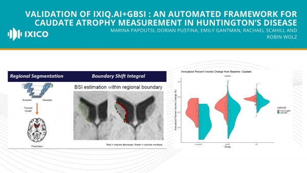
Validation of IXIQ.Ai+gBSI : an automated framework for caudate atrophy measurement in Huntington’s disease
We have developed a fully automated framework that uses deep learning for caudate segmentation (IXIQ. Ai) and generalised BSI (gBSI) for longitudinal measurements. Here, we validate the new method by comparing its volumetric scores with those of the standard manual pipeline (Man+BSI). Man+BSI produced larger caudate volumes than IXIQ.

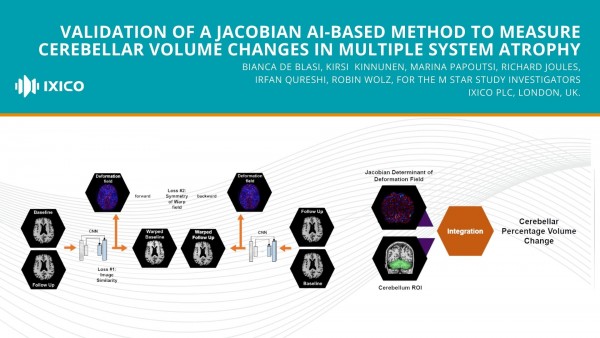
Validation of a Jacobian AI-based method to measure cerebellar volume changes in multiple system atrophy
The accurate, consistent, and scalable estimation of cerebellar atrophy would be highly beneficial for clinical trials in multiple system atrophy (MSA)1-3.
9-16 of 133 results